Introduction
The literature review will focus on the biomechanics of running injuries by reviewing previous publications and research work that have focused on running and the injuries that are caused by running. The biomechanics of running basically refers to the techniques or approaches that form the vital components of running. Having good biomechanics is important for athletes as it prevents injuries or other devastating effects to their physiological foundations. The components that make up the running form include body postures, stride frequencies, foot plant and position, length of strides and foot angle, handgrip position and arm angle swing. Any injuries that affect these component parts lead to faulty biomechanics which in turn make it difficult for a person to run properly (Solkin, 2011).
Literature Review
According to Hawley (2000), there are a growing number of studies and research work that are being conducted on the biomechanics of running with most studies focusing on the forces that act on the body when a person is running. These studies have received a lot of support from technical advances and innovations in the form of speed cameras and marker systems that eliminate the need to manually digitize the frame of the runner when moving. The increasing number of studies that have concentrated on the biomechanics of running have been spurred by the increasing number of people who participate in distance running or jogging (Novacheck, 1998).
Kinetic Movements
Body movements play an important role when it comes to providing some useful insight into how certain changes in the running technique influence the performance of an individual. The study of running provides important information on how a number of biomechanical parameters can predispose an individual runner to various biomechanical injuries (Milner et al, 2006). To gain important information into how runners are exposed to injuries, the review will first focus on the movement of the lower limb segments and the forces that are experienced during each movement of the foot during the running exercise.
According to Tongen and Wunderlich (2011), the forces that are used when running are referred to as kinetics. Kinetics is basically the study of movement and the forces that produce the movement. The study of movement in running is usually measured by a force plate which incorporates Newton’s law of motion. The law basically states that by stepping on the ground, we are producing a vector of force that is both downward and backward which counteracts the force produced by the ground that is generally upward and forward in nature. Ground reaction force is the force known to come from which is mostly deliberate by the force plate. The graph below represents the ground reaction force of a person who is running, speed walking or walking.
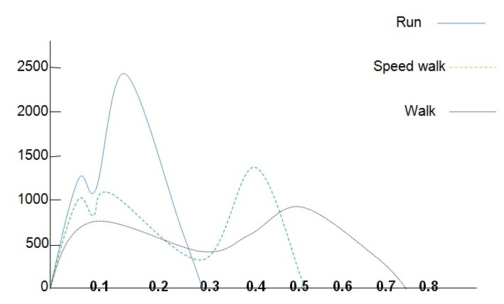
As demonstrated in the graph the ground reaction force for a person who is running is much higher than that of a person who is walking or speed walking. The ground reaction force is an important measure in determining whether a person that is running will suffer from biomechanical injuries. The knee joint contributes to kinetic movement where it is flexed 10̊ as the foot strikes the ground. Once the hip, knee and ankle joints begin to extend during the support period, the foot pushes against the running surface as the body’s centre of gravity accelerates both upward and forward. The ankle joint supports different foot strike patterns which help the runner to continue maintaining contact with the running surface (Hawley, 2000).
The ankle joint is usually flexed by 15̊ to 20̊ dorsiflexion when a person is running. The metatarsophalangeal joint is found between the metatarsal bones of the foot and the proximal bones or phalanges that form the toes. The movements that are allowed by the metatarsophalangeal joints include flexion, extension, adduction and extension. This joint is usually flexed by about 30̊ during the last half of the support phase of running (Taylor et al, 2004). The diagrams below demonstrate the gait cycle of the knee and thigh joints during the running, walking and sprinting exercises.
Biomechanics of Running Injuries
According to Chen (2007), the type of injuries that runners are exposed to include stress fractures, medial tibial stress injuries otherwise referred to as shin splints, chondromalacia patellae injuries and Achilles tendinitis. Chen (2007) identifies the factors that cause running injuries to be excessive training where athletes are subjected to excessive and intensive running, rapid increases of speed during running exercises and stretching of arm and leg muscles; anatomical variables which include the range of motions in the ankles and longitudinal arches and the lower extremity alignment of abnormalities such as the rear foot varus, the Q-angle and the tibia varum.
Biomechanical variables are other factors that lead to injuries in athletes involved in running and these include kinetic variables such as the impact loading rate, intensity of the push off forces and the magnitude of impact forces such as the force plate, knee joint, hip joints and the metatarsophalangeal joints. The hip joint contributes to kinetic running as the hip of a runner is usually flexed between 25̊ to 30̊ as the force plate strikes the ground. The extension of the hip joint usually continues from the toe off at which the point of the hip is in a 20̊ extension (Hawley, 2000).
Novacheck in his 1998 noted that biomechanic injuries usually came about as a result of poor understanding of the pathophysiological biomechanics of chronic running injuries on the part of a runner and trainer. Chronic running injuries usually came about as a result of the repetitive application of small loads of exercises over the course of repetitive running cycles. This contrasted sharply with the chronic injuries that resulted from the sharp distribution of acute traumatic events such as fracture of the ankle or hip joint during physical games such as football or rugby where there is a single large load. Many researchers such as James and Jones, Hill, Fenn and Hubbard who have all conducted studies on the biomechanics of running injuries have attributed these injuries to a variety of intrinsic and extrinsic factors (Novacheck, 1998).
James and Jones in their 1990 study noted that scores of of the running injuries were usually originate from by meticulous anatomic defects that led to precise injury outlines such as the hyperpronation predisposition to posterior tibial syndromes or the genu varum leading to a predisposition of the iliotibial band syndrome. The researchers however noted that such relationships were not found by many researchers to be the major causes of biomechanic running injuries. Most of the biomechanical scholars have contended that making more sense of how and why injuries occur will mostly be achieved by improved diagnostic and counselling activities (James & Jones, 1990).
Cavanagh (1987) who conducted studies on the lower extremity action that was involved in running noted that the abnormal movement of the lower joints over a period of continuous repetitive cycles led to an overuse syndrome due to the increased internal rotation of the tibia. Cavanagh’s 1987 research supported several empirical studies and clinical evaluations that highlighted the fact that shoes otherwise referred to as orthotics that were designed to diminish hyperpronation frequently eliminated painful conditions that came about as a result of running. Roger Mann who is one of the most well known biomechanists to research on running injuries examined the normal and pathological mechanics of hind and mid-foot motions. Mann noted that the sagittal plane ankle motion which usually contributed to most biomechanic injuries was usually accompanied by a rotation of the transverse plane and a rotation of the foot because of the oblique orientation of the ankle joint (Novacheck, 1998).
When a person adopts a gait position, the foot is fixed on the ground which leads to ankle dorsiflexion. This in turn causes the internal rotation of the tibia and pronation of the foot. Through the mitered hinge effect the rotational torques of an individual are usually transmitted to the tibia resulting in the rotational torques along the longitudinal axis. At the initial contact with the ground, the hind-foot is inverted leading to pronation which unlocks the transverse tarsal joint thereby increasing the flexibility of the foot. The transverse tarsal joint acts as a shock absorber when an individual is running, sprinting, speed walking or simply walking (Milner et al, 2007). Any abnormal or excessive pronation on the transverse tarsal joint will lead to a biomechanic injury as demonstrated by the diagram below:
The diagram above represents a schematic view of the difference between normal foot mechanics and hyper-mobile pronated foot biomechanics. Based on the drawing the time to maximum pronation is delayed beyond the 40 percent mark stance. Because of the abnormal excessive pronation that the foot is exposed to, it is not an effective lever of flexibility and movement leading to painful injuries. The transverse tarsal joint is locked and the generation phase has been reached where the foot becomes more rigid (Novacheck, 1998).
Manson et al (2008) conducted an analysis of a homogeneous group of 92 well trained runners so as to determine their biomechanics of running injuries and also establish the relationship between rear foot motion and shock attenuation during running. They examined the athletes for a period of one year so as to determine the rate of injury as a result of running or other exercises that required the use of foot motions. Manson et al (2008) were able to observe the injury rate of the athletes during the one year period to amount to 52 percent. These injuries were mostly in the form of stress fractures, shin splints and Achilles heels which affected the lower leg of most of the athletes. The number of athletes identified to have shin splints and stress fractures represented 22.7 percent of the population under observation (Manson et al, 2008).
The results of their study revealed that there was a negative correlation between the peak tibial acceleration and the magnitude of the rear foot motion based on injuries that occurred on various locations within the foot (r2=0.35-0.65). This negative correlation meant that objective biomechanical parameters were implicated in running injuries or they conversely led to biomechanics running injuries. Manson et al (2008) noted that gait as a predictor of various types of running injury was important when it came to preventing biomechanics injuries during running or other exercises that required foot movement. The predetermined biomechanical parameters proved to be important when it came to designing suitable shoes that would be used by runners so as to prevent biomechanics injuries. Manson et al’s observations and conclusions coincided with those of researchers like Pink and Jobe who summarized their observations on the importance of having the right shoe to prevent biomechanic injuries (Novacheck, 1998).
Bates et al concluded that there was no one shoe that was suitable for runners and that shoe companies had to design shoes that would manage shock absorption and also offer stabilization to the runner. Many shoe designers have focused on the attenuation of the shock of heel strike when designing their shoes as well as the control of the hind foot motion during the loading response. Well constructed shoes provide appropriate shock absorption and the stabilization of the foot reducing the load placed on the transverse tarsal joint. According to Cook et al (1985) the intrinsic factors of each individual runner such as the degree of pronation, flexibility of the foot and their body weight were all important factors that needed to be considered by a runner when selecting an appropriate shoe.
Based on Cook et al (1985), the mechanical testing of shoes for shock attenuation showed that 75 percent of the shock attenuating capabilities of a shoe were retained after 50 miles and 67 percent after 100 to 150 miles. However, despite such advances in shoe wear, the rate of injury amongst distance runners continued to increase significantly which lent credence to the fact that chronic injury patterns among runners were caused by other forces apart from the shock of the initial contact of the foot and the alignment stance in the shoe wear.
Conclusion
The literature review has focused on the various studies and research work that have been conducted on the biomechanics of running injuries. The research has focused on running and it has identified the various biomechanics injuries that runners are prone to when they are running. The major findings that can be generated from the research are that individual runners suffer from individual injuries which mean that developing a one for all solution for biomechanics running injuries will be an impossible task. While the review has been able to identify common injuries that affect majority runners, developing suitable solutions to deal with biomechanics problems has eluded most of the researchers with some concluding that the development of a flexible shoe will reduce the number of injuries that affect runners. These research findings can be contributory to the existing literature and research work that has been done on biomechanics of running injuries as it has been able to highlight the various components that come into play when a person is running.
References
Cavanagh, P.R., (1987). The biomechanics of lower extremity action in distance running. Foot Ankle, 7(4): 197-217
Chen, H.C., (2007). Biomechanical aspects of running injuries. Web.
Cook, S.D., Kester, M.A., Brunet, M.E., & Haddad, R.J., (1985). Biomechanics of running shoe performance. Clinical Sports Medicine, 4(4):619-626
Hawley, J.A., (2000). Handbook of sports medicine and science: running. Oxford, UK: Blackwell Science Limited
James, S.L., & Jones, D.C., (1990). Biomechanical aspects of distance running injuries. New York: Human Kinetics Books
Manson, N.A., McKean, K.A., & Stanish, W.D., (2008). The biomechanics of running injuries. Journal of Bone and Joint Surgery, 90(52):
Milner, C.E., Davis, I.S., & Hamill, J., (2006). Free moment as a predictor of tibial stress fracture in distance runners. Journal of Biomechanics, 39: 2819-2825
Milner, C.E., Hamill, J., & Davis, I., (2007). Are knee mechanics during early stance related to tibial stress fracture in runners? Clinical Biomechanics, 22: 697-703
Novacheck, T.F., (1998). The biomechanics of running. Gait and Posture, 7:77-75
Solkin, M., (2011). The biomechanics of running. Web.
Taylor, D.T., Sage, R.A., & Pinzur, M.S., (2004). Arthrodesis of the first metatarsophalangeal joint. American Journal of Orthopedics. 33 (6): 285-288
Tongen, A., & Wunderlich, R.E., (2011). Biomechanics of running and walking. Madison, US: James Madison University